The J.Stefan Institute
home page is located at http://ijs.muzej.si/
The F-2 home page is located at http://rubin.ijs.muzej.si/
 Microanalytical Center
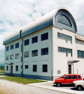
Microanalytical Center (MIC) is located at J. Stefan Institute Reactor
Center at Brinje. It specialises in Ion Beam Analytical (IBA) methods
and research in experimental atomic physics. It performs quality assurance oriented
studies with:
 Particle Induced
X-ray emission (PIXE)
Particle Induced
X-ray emission (PIXE)
-
Particle Induced gamma-ray emission (PIGE)
-
Nuclear Reaction
Analysis (NRA)
-
Rutherford Backscattering
Spectrometry (RBS)
- Forward Scattering
Spectrometry (FSS)
-
Elastic Recoil Detection
Analysis (ERDA)
- Auger Electron Spectrometry
(AES)
- High Resolution
X-ray Spectrometry (HRXS)

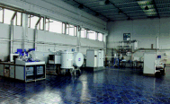
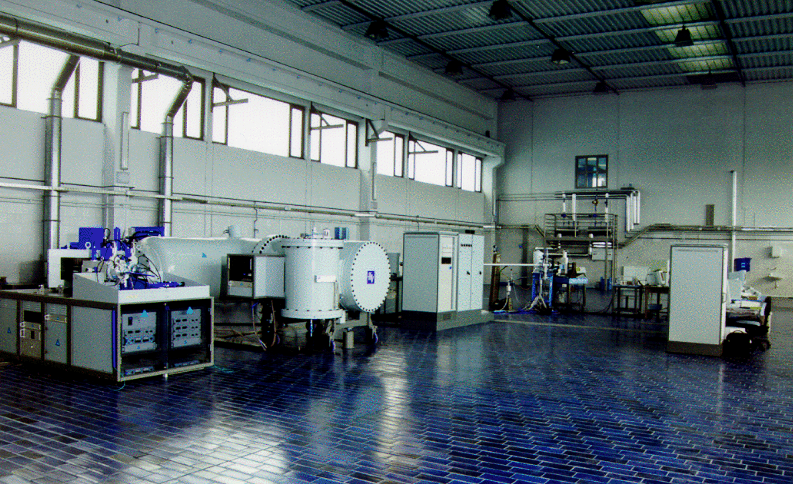
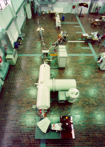
Tandetron: The new 2MV tandem accelerator installed with the financial
aid from IAEA and Ministry for Science and Technology.

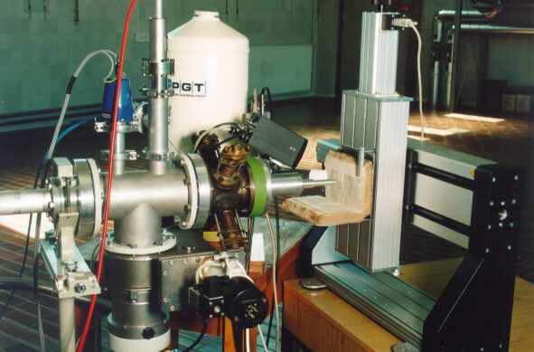
Tandetron: In-air IBA beamline.
Head Milos Budnar
![[Photo of group]](../images/sk_icon.jpg)
Research staff from the right to the left:
 Milos
Budnar
Milos
Budnar
- Zdravko
Rupnik
- Matjaz
Zitnik
- Ziga
Smit
- Benjamin
Zorko
- Franz
Gasser
- Primoz
Pelicon
- Matjaz
Kavcic
- Artur
Mühleisen
- Marjan Ravnikar
For collaboration or further
information contact Milos Budnar
MAIN RESEARCH FACILITIES
- HVEE TANDETRON accelerator
with 2MV terminal voltage, duoplasmatron and sputtering ion sources, switching
magnet with five ports for beamlines, two beamlines installed:
 PIXE/PIGE/NRA beamline
in air IBA beamline
- 1.5MV Van de
Graaff accelerator (located at J. Stefan Institute main site)
RESEARCH ACTIVITIES
- Basic research in
atomic physics
- atomic single and
multiple ionisation by charged particles
post-collision
interaction (PCI)
hypersatellites
in gaseous targets
second order radiative
effects in X-ray spectra
models of inner
shell ionisation and decay after charged particle or photon impact
- Education
- PIXE
excercise for physics students
experimental atomic
physics and interdisciplinary subjects for M.Sc. and Ph.D. degrees
public information
about the safe use of low-energy accelerators for research and industry
specialisation
programs for researchers from industry
international training
courses
- Interdisciplinary
research
- studies
of aerosols from urbane environment
occupational health
studies of aerosols from working environment
determination of
hydrogen concentration profiles in material surfaces
depth profile determination
of tool hard coatings
thin film analysis
usewear studies
of prehistoric tools
elemental contents
in antic metal alloys
DEVELOPMENT OF
 accelerator-based
spectroscopic methods (IBA methods)
accelerator-based
spectroscopic methods (IBA methods) -
electron spectroscopies
- high-resolution
X-ray spectroscopy
-
vacuum systems for beam transport and measurements
-
data acquisition and processing hardware and software
INTERNATIONAL COLLABORATION
 Johannes Kepler University, Linz,
Austria
Johannes Kepler University, Linz,
Austria - University
of Fribourg, Fribourg,
Swiss
- ATOMKI,
Debrecen, Hungary
- University of Rome III, Rome,
Italy
- ELETTRA,
Trieste,
Italy
- University of Singapore, Singapore
- University of North Texas,
Denton, USA
- University of Oxford,
Oxford, Great Britain
- University Pierre and
Marie Curie, Paris, France
Oxford, Great Britain
RESEARCH ACHIEVEMENTS IN 1996 AND 1997
Multiparticle excitations and many body decay processes in inner atomic shells are subject
of numerous studies in experimental atomic physics.
In order to study second and higher order atomic excitations, we have
collaborated in measuring Ne and Ar photoabsorption spectra on the VUV
undulator beamline at
ELETTRA,
Trieste. Some doubly excited states were observed
for the first time. Of equal interest are X-ray transitions in single or
multi-ionised atoms,
where many-body decays occur in addition to dominant diagram transitions.
Here, we have measured
in collaboration with the University
of Fribourg, and PSI, Villingen,
the K-LM and K-MM processes of the radiative Auger effect
(RAE) in Ar, Kr and Xe for the first time.
Additionally, theoretical treatment
of RAE has led to realisation that it is necessary to consider the variation of the
atomic potential during the decay in order to achieve a better physical model.
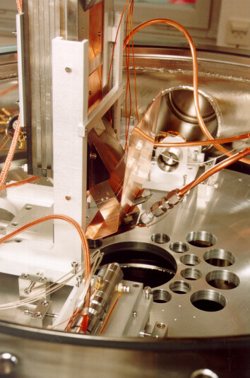
The new Auger electron gas spectrometer has become operational. It
enables research on nonradiative atomic transitions and ionisation
processes (for example, it was used for measuring hypersatellites in
Ne). It is equiped with magnetic field shielded UHV chamber,
electrostatic energy analyser, electron gun and VME based electronics.
It is run with dedicated home developed software.

AES: Inside of the Auger electron gas spectrometer
The new tandem electrostatic accelerator (TANDETRON) has been installed and
two new beamlines constructed,
with the financial support from IAEA and the Slovene Ministry for Science and Technology.
The accelerator facility can provide improved research possibilities
for investigation in several research fields related to atomic
physics: chemistry, biomedicine,
new materials, environment, archaeometry and others.
We have continued with the development of new spectroscopic methods
with accelerated ions (IBA methods) and X-rays. The feasibilities of the
ERDA have been tested on the standard samples. We have tested their feasibility
on a number of interdisciplinary fields.
 RELATED DOCUMENTS
 Research
projects
Research
projects - International
agreements
- Publications
DESCRIPTION OF SOME IBA METHODS
![[IBA methods]](../images/iba_ic.gif)
Figure: Schematic presentation of different IBA methods.
Particle Induced X-ray Emission
Light projectiles (protons or He ions) with the energy of few MeV
excite the atoms by knocking out electrons from their inner shells (K, L).
Vacancies produced by such processes are filled with electrons from the
outer shells. The available energy of the transitions is manifested in
the emission of characteristic X-rays (K, L-lines). In the spectrum areas
under the peaks belonging to the characteristic lines are proportional to the
average elemental concentrations of different elements in the sample.
Characteristics of the PIXE method:
-
Protons or He ions with the energy of a few MeV are
usually used as the projectiles
-
Ion beam with the area cross section from
microm2 (micro PIXE) to a few mm2 and the current densities from 1 pA up to
100 nA per mm2 are applied
-
Concentrations of the elements from C up to
U can be measured simultaneously
-
Trace elemental concentrations below ppm
can be determined in the samples
-
In-air analysis is often applied for studies
of special samples
Use:
-
Trace and major element analysis
-
Various samples
Nuclear Reaction Analysis
Nuclear Reaction Analysis (NRA) is based on the nuclear reaction between
the projectile and the target nuclei. The reaction cross section depends
on the projectile energy and on the counterparts involved in the collision.
If the projectile has sufficient energy to penetrate the Coulomb's barrier
of the target nucleus its energy is imparted to the nucleons. An intermediate
nucleus in excited state is formed which decays either by photon or particle
emission. Measuring the yields of reaction products when changing the projectile
incident energy the elemental concentration depth distribution can be obtained
also.
Characteristics of the NRA method:
-
Different ions with various energies
are used as the projectiles
-
The ion beams with the cross area of 1mm2 and
the current density up to 1 A/mm2 are usually applied
-
The elemental concentration
sensitivity for some elements is high due to large cross sections for particular
reactions
-
One element concentration depth profile can be measured during
the measurement time
-
The depth resolution of the profile in the near surface
region is below 10 nm
-
Usually gamma rays with the energies of several MeV
as well as the light particles (p,n,d,alpha) are detected
Use:
-
Elemental analysis, element concentration profiles
-
Various samples
Rutherford Backscattering Spectrometry
In the Rutherford Backscattering spectrometry (RBS) large angle scattering
of the incident particles by the target nuclei is employed. At fixed detector
angle the energy of the projectiles emerging from a given depth determines
the mass of constituent atoms and their location. The number of counts
is proportional to the atomic concentration of the element. The projectiles
arriving from different depth have different kinetic energies due to the
slowing down in the target. The spectrum given as the number of recorded
scattered projectiles per energy interval serves for the evaluation of
the elemental depth distribution.
Characteristics of the RBS method:
-
Protons,
He or Li ions with the energy of few MeV are usually used as projectiles
-
Projectiles scattered by the target atoms are detected at the angle near
180 degree with respect to the incident beam direction
-
Typically the ion
beams with the cross area of 1mm2 and the current
density up to 100 nA/mm2
are applied
-
The elemental concentration sensitivity: high for Al to
U (< 0.1 at.%), low for Li to Mg (< 10 at.%), insensitive
for hydrogen
-
Depth concentration profiles of the elements heavier than
the projectile are measured simultaneously
-
Depth resolution of the
depth profile in the near surface region is 15-20 nm
Use:
-
Elemental concentration profiles
-
Thin layer analysis, hard coatings studies
Elastic Recoil Detection Analysis
Elastic Recoil Detection Analysis (ERDA) is based on recording nuclei
which are knocked out by the incident projectiles. In order to detect the
recoiled nuclei the target is tilted at the grazing angle with respect
to the direction of the incident beam. Since the impinging projectiles
are scattered in all directions an absorber attached to the detector window
is used to discriminate the recoiled particles from the scattered ones.
A thin foil made from Al or C, which stops the projectiles and let through
the recoiled nuclei only, is used. By measuring the energies of the recoiled
nuclei coming from a particular depth and counting their yields a depth
profile of the element is obtained.
Characteristics of the ERDA method:
-
He, Li or heavier ions with the energy of few MeV are usually used as
projectiles
-
The target tilting angle is 10 to 20 degrees with respect to the incident
beam
-
The recoiled nuclei are detected at fixed detection angle between
10 and 40 degrees
-
The ion beams with the cross area of 1mm2 and the current
density of up to 100 nA/mm2 are usually applied
-
Depth concentration profiles
of the elements lighter than the projectile are measured simultaneously
-
Depth resolution of the hydrogen concentration profile in the near surface
region is estimated to be from 25 to 50 nm when helium ions with the energy
of 1 MeV are used
-
Sensitivity for hydrogen concentration
determination is below 0.1 at.%
Use:
-
Hydrogen and light element concentration profiles in near surface
region
-
Material studies, material surface analysis
Last update January 23, 1998; am, dgc
![[Photo of group]](../images/sk.jpg)
![[IBA methods]](../images/iba.gif)